CLAS Faculty Earn College Awards For Teaching Excellence
Shown (from left) are Paola Lopez-Duarte, Lennin Caro, Interim Dean John Smail and Samantha Suptela.
For their exceptional teaching and student engagement, Paola Lopez-Duarte, Samantha Suptela and Lennin Caro have received the College of Liberal Arts & Sciences’ 2023 Excellence in Teaching Awards. Six other faculty were honored as finalists for three awards.
Lopez-Duarte, an assistant professor in the Department of Biological Sciences, received the Integration of Undergraduate Teaching and Research Award. Suptela, an assistant teaching professor in the Department of Biological Sciences, received the Award for Outstanding Teaching by a Full-Time Lecturer. Caro, a faculty member in the Department of Anthropology, earned the Award for Outstanding Teaching by a Part-Time Faculty Member.
The college recognized the honorees at an awards ceremony and reception on May 3. In addition to the award recipients, finalists who were honored are:
- Loc Nguyen, Mathematics and Statistics; and Tina Shull, History: Integration of Undergraduate Teaching and Research Award
- Jesse McKee, Criminal Justice and Criminology; Erin Godly-Reynolds ’20 Ph.D., Psychological Science; and Scott Wilde, Mathematics and Statistics: Outstanding Teaching by a Full-Time Lecturer
- Jane Walsh ’13, ’19 M.S., Biological Sciences: Outstanding Teaching by a Part-Time Faculty Member
Paola Lopez-Duarte
Paola Lopez-Duarte has made it her mission “to build inclusive research communities and make the process of scientific discovery accessible to all.” Lopez-Duarte develops undergraduate and graduate courses in Marine Ecology and Marine Sciences that help students build skills to think like a scientist through courses that integrate research methods, experimental design and data interpretation. She has implemented unique assessment methods and a highly interactive community of undergraduate students to dig into primary literature and various research methods.
Lopez-Duarte also developed a “Meet My Marine Scientist” assignment where students contact scientists they are interested in working with or with whom they share an identity. Lopez-Duarte is dedicated to participation in the Transforming STEM Academy, where she uses new tools from the learning community to help her classrooms feel more navigable to students.
She has mentored many students through the Office of Undergraduate Students Scholars program and the Honors program. Undergraduate research includes laboratory and field work where students travel to sites to collect and process samples and use analytical tools. She stresses the importance of hands-on learning activities that task students, for example to find research articles in databases and develop annotated bibliographies. Her students present at regional and national conferences, and she routinely publishes original work with undergraduates as co-authors.
She serves as a faculty advisor for the Society for Advancement of Chicanos/Hispanics and Native Americans in Science (SACNAS), a chapter she initiated in the department. She has nominated students for Sigma Xi, the Scientific Research Honor Society, and supported the students to apply for Sigma Xi’s “Grants-in-Aid of Research” and attend an annual conference.
Samantha Suptela
Samantha Suptela, ’03, ’12 Ph.D., joined the Charlotte faculty as a full-time lecturer in fall 2018 and is now an assistant teaching professor with expertise in immunology and microbiology, with research focused on host responses to bacterial infections of bone and brain cells and equity in STEM education. Along with her Charlotte degrees, Suptela earned a master’s in public health from the University of Virginia in 2013.
She is currently working toward a master’s of education in learning, design, and technology, with a concentration in online learning and teaching and has recently earned the “Essentials of Teaching & Learning” certificate. She has won multiple grants to improve her courses, and she was a Faculty Pioneer in the Student Experience Project, an initiative that seeks to dismantle inequities in college success by transforming the student experience. As a member of The Center for Teaching and Learning’s “Active Learning Academy,” she has learned many ways to engage students through innovative teaching techniques that create a supportive learning environment.
One student commented, “I LOVE the classroom environment that Dr. Sam creates. She makes me feel so comfortable.” Another said, “I felt very confident in the course and in my chances to succeed regardless of my race and gender. I felt that I belonged and I never felt that I was being treated as if I did not belong.”
In 2019 Suptela was nominated for the Alpha Chi Omega Professor of the Year Award and the UNC Charlotte Student Government Association Professor of the Year Award. Suptela has set her goal as being “someone who inspires future generations of scientists and educators — making a difference in their lives and in the future of the discipline as a whole.”
Lennin Caro
Lennin Caro, ’14, ’17 M.A., received bachelor’s and master’s degrees in anthropology from UNC Charlotte, focusing his graduate work on the different ways evangelical Christian groups at UNC Charlotte structure their outreach and mission programs to fellow students and more widely in the community. Caro is known for the sophistication of his analysis of field data using conceptual frameworks from anthropological work on religion and on economic culture.
Caro began teaching at Charlotte in the fall 2018 semester. Soon after he began teaching, Caro became a full-time research the Camino Research Institute, the research arm of a community organization in Charlotte that works with local Latino people. Caro has used his native language skills and his anthropological training in his current role as lead community researcher, generating and disseminating data on the social and health needs of Latino families in Charlotte.
Over 700 students have taken Lennin’s classes, including Introduction to Anthropology, Western History & Culture, Foundations of Anthropological Theory and Themes in Sociocultural Anthropology. His students have benefited from his skill as a teacher and his experience as a full-time community researcher. His deep engagement with the material and with his students is outstanding and is demonstrated in his teaching evaluations.
“If this campus had more professors like Mr. Caro, every student would be able to understand difficult theories and concepts, learn in a stress free environment, and would always feel respected,” a former student wrote. “Mr. Caro is the epitome of what a teacher is and deserves acknowledgment for his approach, his capabilities, and his successful methods.”
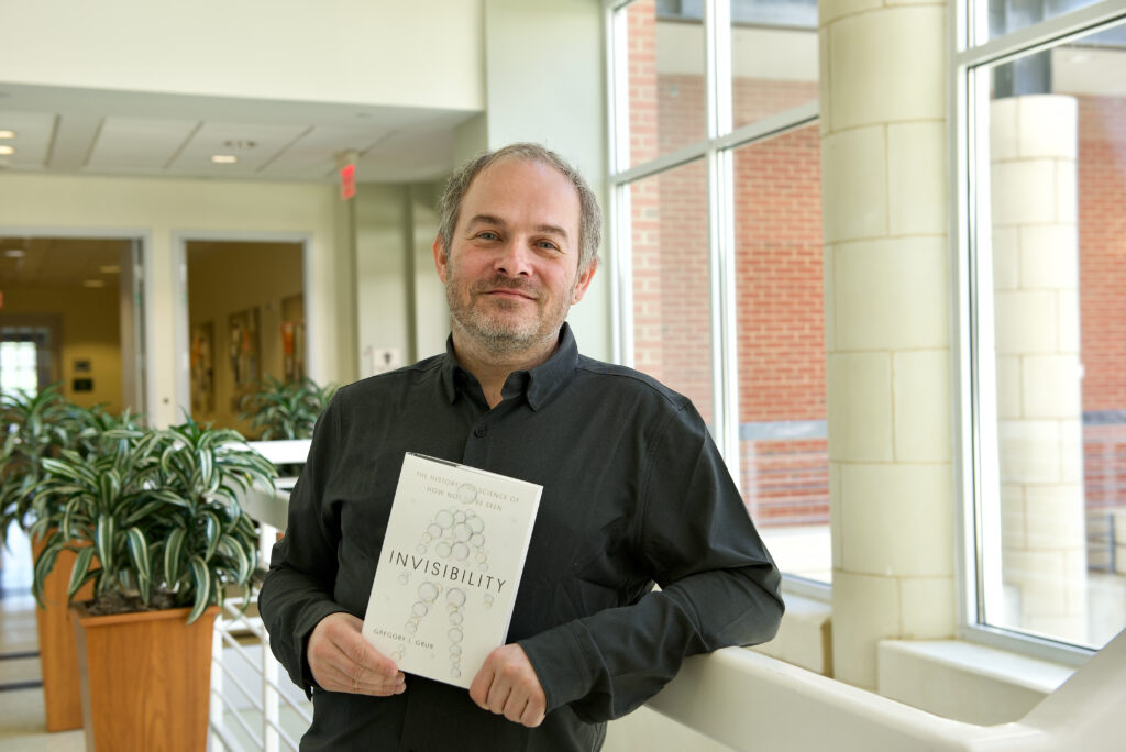
New Book “Invisibility” Brings Continued National Visibility For Charlotte Optical Physicist Gregory J. Gbur
Charlotte optical physicist Gregory J. Gbur turned to stacks of science fiction stories, a treasure trove of horror films, and his scientific expertise and discoveries when writing his new book, “Invisibility: The History and Science of How Not to Be Seen.”
“Invisibility” is not a purely scientific book, although certainly Gbur has written his share of those. This is an entertaining book for readers curious about one of the most popular plot devices from science fiction and an understandable discussion of the scientific concepts that one day might transform these tales into truth.
“There is a long history in both science and science fiction of people trying to imagine how things might be invisible or how invisibility might be related to interesting phenomenon,” said Gbur, who arguably is the world’s leading expert on invisibility fiction.
“My book looks at the earliest science fiction stories about invisibility from 1859 all the way to modern-day research as people today try to figure out how we can make an invisibility cloak and what other strange phenomena we can do,” Gbur said. “The book is really made up of the stories of what people were thinking at the time they were making various discoveries, how they were making them, and the troubles they had.”
Gbur, named an international Optica Fellow for his significant research in coherence theory, singular optics and the intersection of these disciplines, studies invisibility and related phenomenon as one aspect of his diverse research agenda.
“I still in fact do some research on invisibility,” he said. “I have an academic paper out now for review that talks about things that I write about in “Invisibility.” My Ph.D. work, which I completed in 2001, was on invisibility-related stuff. The main academic papers that caused invisibility cloaks to become a popular research topic didn’t come out until 2006. So, I say that I was doing invisibility before it was cool, as kind of a hipster invisibility theorist, and also, because of my background, I had a front row seat to all the discussions that were happening about invisibility.”
A Publishers Weekly Top 10 Science Book, “Invisibility” was reviewed in The New York Times and featured as a “Must Read” book by the Next Big Idea Club. Gbur recorded a short talk about the book for the Next Big Idea Club, and site curators Susan Cain, Malcolm Gladwell, Adam Grant and Daniel Pink will include it in a book list they will narrow to a handful of finalists and, ultimately, to two official season selections.
Gbur also joined renowned astrophysicist and science communicator Neil deGrasse Tyson and guest host and comedian Negin Farsad for a StarTalk appearance with a lively discussion of transparency versus invisibility, how metamaterials help us interact with different wavelengths, what light has to do to make something invisible and other topics.
Gbur’s book is geared particularly for readers with interests in science, history and science fiction, with a writing style honed through Gbur’s posts on his two blogs on horror fiction, physics, and nature, including Skulls in the Stars. “Invisibility is written without equations and without extremely technical descriptions,” he said. “I really try to explain things in understandable terms, embracing the weirdness in both science and science fiction, and I try to keep it fun.”
Fans will note that the book subtitle pays tribute to a classic sketch by British comedy troupe Monty Python, and in addition to the standard bibliography, it also features an invisibibliography, both signaling that this volume is not meant to be a boring slog of a read.
Gbur’s first book for a non-scientific audience, “Falling Felines and the Fundamentals of Physics,” also published by Yale University Press, explored the science of falling cats righting themselves as they land on their feet and related phenomenon. Gbur, who has fostered more than 20 cats and currently has 5 of his own, had set aside the intended book about invisibility when he was drawn to the cat topic. He made sure to include a historic photograph related to cats and invisibility in his new publication, and he’s decided that every future book will feature a cat illustration.
Here are a few more thoughts from Gbur about invisibility and related topics.
Do you think invisibility will be possible in the future, or do you think it will remain just part of science fiction?
It is always hard to predict the future in science, so we should recognize that new information can always change things. I imagine we will see some remarkable demonstrations of near-invisibility in the future, though I suspect that these will be primarily tech demonstrations and not products put to use for sinister purposes or otherwise. The construction and design of invisibility devices seems too challenging, and thus probably cost prohibitive. I do believe that the insights that we have learned from the theory of invisibility, and the advances that come from studying the possibility of invisibility, will have many benefits to optical technology in the future — even if we cannot see them directly.
How does science fiction interact with the science of invisibility?
It is fascinating to go back and look at science fiction stories about invisibility. There are way more of these than people realize. I was myself surprised by how many science fiction invisibility stories I found that nobody talks about any more. People writing about invisibility in science fiction felt they needed to at least justify it somehow using the science that was available at the time. The science fiction gives this sort of snapshot of the science of its era, including optics.
H.G. Wells, when he wrote “The Invisible Man,” knew enough optics to try to explain the optics side of whether an object could be invisible. But he also had to try and explain how might one achieve invisibility. He drew upon the fact that X-rays had been discovered only a couple of years earlier, and everybody was confused by X-rays. They saw X-rays as a form of invisibility because there were images where a person was see-through. There were newspaper articles about people selling X-ray-proof underwear. People were afraid they were going to be spied on when they were walking down the street.
They also at times got it pretty close to how we now imagine invisibility cloaks to work. There were a number of authors that ended up using the same analogies that the modern invisibility cloak people ended up using to explain how it works.
How did the scientific research you do influence the book?
As I was writing the book, it evolved not just into the story of invisibility, but it ended up being closely tied to the history of optics itself and how our understanding of light has evolved. I hope that when people read this, they’ll gain a better understanding of light and how light works. That is something that most people don’t get a whole lot of exposure to in school. Basic physics classes talk just a little bit about light and optics and how it works. Even a lot of physicists don’t delve very deeply into optics. So, part of my hope is just to make the whole science of light a bit more accessible, and in a fun way by talking about how our understanding of light has also driven our understanding of how invisibility might work.
Why does understanding optical science, at least to a certain degree, matter, and why do you describe the field as beautiful?
It matters if we understand optics simply because it’s a beautiful field of study, and not a lot of people understand it. It’s also worth understanding because so much of our modern technology is based on optics. For example, our Internet communications, our phone communications, often are transmitted through fiber optic cables, which all really depend on a deep understanding of optics.
Optics is beautiful partly because of the images we can make of some of the phenomena. Everybody has seen rainbow patterns and interference patterns when you mix different light waves together, especially colored light waves. There are so many complicated phenomena that produce these elegant images.
For me also, a lot of the beauty in optics is just how well the subject works and how well it all fits together. It’s a mature enough field now that we have this deep understanding of it, and when you make these connections of why things work the way they do, it’s lovely to see it all come together.
What lies ahead with the science?
There have been so many unusual and interesting discoveries about how light interacts with matter, and there is a possibility that much more of our society may change and be transformed by these recent discoveries that are connected somewhat to invisibility. It’s not that we’re going to have people walking around in invisibility cloaks but that the knowledge that we’ve learned from studying invisiblity may turn into more interesting things.
Modern advances in optics have changed the way we look at things like cameras and traditional lenses, as just one example, like with our Center for Freeform Optics at UNC Charlotte. A lot of the current discussion is around metamaterials, or the idea that you can design materials that have optical properties that are not found in nature.
Amazing optics being done now may transform our society. I hope this book will give people a little bit of an understanding of where that may take us.
Words and Images: Lynn Roberson
Chemistry in the news: Featuring Dr. Vivero-Escoto
Dr. Juan Vivero-Escoto was recently interviewed by WBTV on your side about his research on antibiotic resistant bacteria. Check out the segment here to learn more about the amazing research being done at UNC Charlotte!
Dr. Walter: Recipient of the 2022 CLAS Teaching & Research Integration
Michael Walter joined the faculty at UNC Charlotte in 2011. Since then, he has secured over $2.5 million in external grant funding and published two dozen papers. He is the inventor of co-inventor for three patents, one of the patents is licensed to a local company.
His research program, and his real-world teaching illustrations, are built around the study of various materials that use light interactions for energy. Students learn how powerful organic chemistry photochemical tools can be used to address scientific challenges.
Undergraduate students are attracted to Walter’s research laboratory as early as their sophomore year as a result of their experiences in his classes. He has mentored 53 students from a variety of majors, including chemistry, biology, physics, public health, and mathematics.
His interactions with undergraduate students have resulted in over 50 research talks and posters with several invited talks, all presented by the students.
He developed an “e-molecules” activity where students research the structure of widely used organic molecules that might be used in pharmaceuticals or other materials. The course also includes a hands-on photochemistry activity, called “Juice-from-Juice” where students build blackberry juice, dye-sensitized solar cells. The activity is a powerful example of how an organic molecule extracted from blackberry juice can be used to harness sunlight and convert it to usable electricity and power.
A new activity in his classes looks at the connections among the luminescent materials encountered in daily lives, from organic light-emitting diodes (OLEDs) in cell phone screens to the bioluminescence from fireflies seen on a summer night.
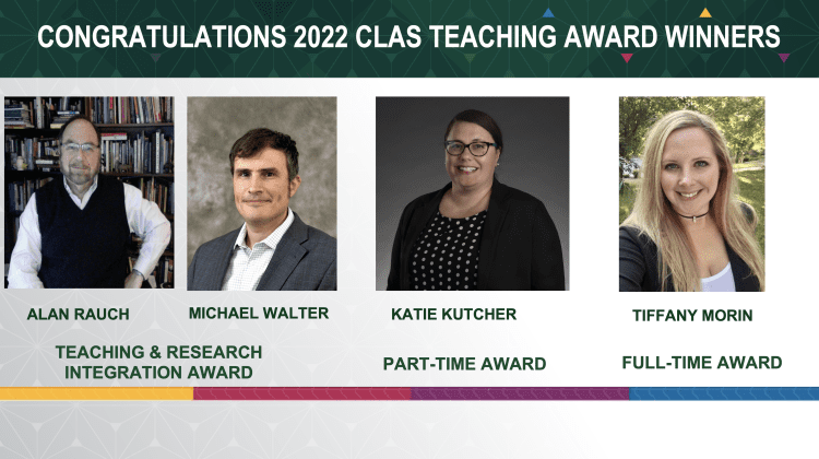
Michael Walter was the recipient of the 2022 CLAS Teaching & Research Integration award
Inaugural Distinguished Leadership Awards Recognize Five Honorees Who Have Helped Make CLAS Strong
The five inaugural recipients of the College of Liberal Arts & Sciences (CLAS) Distinguished Leadership Awards are “impeccable people” who have never let the college or UNC Charlotte down, emcee Ohavia Phillips ’15 told the family members, friends and colleagues who honored them on March 23.
CLAS Interim Dean John Smail agreed. “It takes leadership, and it takes people who are willing to commit time and energy,” Smail said. “It takes vision and belief to make this a top-tier institution. It’s with great pride and great humility and gratitude that I recognize our honorees.”
The new awards, to be presented each academic year, recognizes the achievements of students, alumni, faculty, staff and supporters who have made the College strong. Inaugural honorees who were recognized at a celebration at the Harris Alumni Center at Johnson Glen are:
- Twig Branch ’11 M.A., ’16 M.A., ’18 M.A.
- Nancy A. Gutierrez, Dean Emerita
- Margaret Kocherga ’16, ’20 Ph.D.
- Shawn Long, former Senior Associate Dean, Honored Posthumously
- Boris “Bluz” Rogers ’06
Among special guests were family members and friends of Long, who served Charlotte and the college as a faculty member and administrator for almost two decades. Long died Jan. 14, 2021 following a serious illness. He had joined Kennesaw State University in Georgia as dean of the College of Humanities and Social Sciences in July 2019.
Others recognizing the honorees included Provost and Vice Chancellor for Academic Affairs Alicia Bertone and Andrew Baker ‘15, president of the college Alumni Council.

Margaret Kocherga
Kocherga is an award-winning early-stage entrepreneur, scientist, TEDx speaker and choreographer who has been featured in Bloomberg, The Business Journals, WSOCTV, Yahoo, and Charlotte Magazine. Originally from Ukraine, she finds joy in exploring other cultures and has visited 32 countries. She is the founder and CEO of Light and Charge Solutions, LLC and Margik Inc., which were created with a focus on the commercialization of organic electronics materials and Organic LEDs (OLEDs).
Boris “Bluz” Rogers
Rogers is an Emmy-award winning poet, director of creative engagement for Blumenthal Performing Arts and the coach of the three-time National Poetry slam Championship team, Slam Charlotte. The author of three books is also a recording artist, and his voice is a progressive tool used to foster transformation. Rogers has worked with organizations including CBS Radio, ESPN and SPEED TV and was the first performer at the opening ceremony of the NASCAR Hall of Fame.
Twig Branch
Branch worked as part of his family’s business before moving to Charlotte and retiring. He then obtained master’s degrees from Charlotte in religious studies, history and Latin American studies. Branch has helped raise nearly $5 million to support CLAS and helped establish initiatives including the Salon Outreach Program, Witness in Residence Program, Equity in Memory and Memorial Project, Pharr Buchenau Study Abroad Fund, Ruff English Scholarship, several travel grants and Atkins OutLoud.
Shawn Long, In Memorium
Long began his career at Charlotte, where he held faculty and administrative roles, including as CLAS senior associate dean. In 2019, he became the first black dean at Kennesaw State University. He received many awards in his lifetime, including the Lyman T. Johnson Torch of Excellence from the University of Kentucky’s African American Alumni. Long’s final career achievement was facilitating a $9 million gift to name the Norman J. Radow College of Humanities and Social Sciences.
Nancy A. Gutierrez Dean Emerita
Gutierrez retired in June 2022 after 17 years as CLAS dean. She led efforts to create new academic departments and centers, including the Office of Interdisciplinary Studies. She was instrumental to establishing a Phi Beta Kappa chapter and development of the LEADS applied learning initiative. Gutierrez served as president of the Council of Colleges of Arts and Sciences and offered six years of service to North Carolina Humanities, including as Board chair from 2019 to 2021.
Read More About The Honorees
Pictured (left to right) Twig Branch, Nancy A. Gutierrez, Shawn Long’s aunt Lanora F. Long, Margaret Kocherga, Boris “Bluz” Rogers and Andrew Baker | Image by: Kat Lawrence
Jacob Horger selected to present at Faculty Showcase
Congratulations to Dr. Jacob Horger, Teaching Assistant Professor in the Department of Chemistry. Dr. Horger has been selected to participate in the 5th annual Faculty Showcase. The committee in charge of the showcase said of Jacob’s work, “The quality of the work he recently undertook with the Office of Assessment and Accreditation’s Scholarship of Assessment program to use Gradescope as a way to provide quick, high-quality feedback to students and to monitor the impact of pedagogical changes in his course deserves special recognition, and we believe his efforts will assist faculty across campus to develop their own teaching.” Faculty Showcase Announcement
Please join us for this year’s Faculty Professional Development Showcase and learn how fellow faculty made improvements in their teaching as a result of their participation in professional development activities around campus. The purpose of this event is to raise awareness and celebrate faculty best practices at UNC Charlotte.
This year’s event will take place on the UNC Charlotte campus on April 6, 2023 from 10:30 AM – 12:30 PM in the Halton Reading Room located in Atkins Library. A light lunch will be served.
This Showcase is hosted by the Office for Assessment and Accreditation, Center for Teaching and Learning, Atkins Library, and Communication Across the Curriculum.
Please register online by Friday, March 24th to let us know you’ll be attending. Visit the Faculty Showcase webpage on the Office of Assessment & Accreditation website to learn more about the upcoming showcase and past events.
Non-Traditional Students Benefit From Scholarships, Other Support Through OASES
U.S. Coast Guard veteran Zach Bessant is achieving his goals through the James William Kirk Scholarship established by Kim Kirk Steffan.
Bessant, a junior physics major, was one of two speakers at the recent OASES scholarship luncheon. Toni Moss, a junior pre-communication studies major who also is a scholarship recipient, was the second speaker. Both have benefitted from life-changing opportunities presented through OASES.
After high school graduation in 2007, Bessant joined the Coast Guard and was stationed at various installations across the United States, concluding his service in Wilmington, N.C. He enrolled at Cape Fear Community College to study machining. He later transferred to Charlotte, following treatment for cancer. At Charlotte, he said he is inspired by the concepts of applied math and physics and the possibilities of working in the space industry or starting his own state-of-the-art machine shop.
Moss, a mother of two, would like to open her own business and establish a nonprofit mentorship program for underprivileged adults who aspire to work in human resources, public relations or a similar field. In 2020, she completed an associate degree in hospitality management from Central Piedmont Community College. Most recently, she worked as an HR coordinator for Raytheon Technologies through a staffing agency, and then enrolled at Charlotte, where she has received scholarships through the WINGS and ANSWER programs.
OASES, the Office of Adult Students and Extended Services, is dedicated to meeting the unique needs of nontraditional students. Each academic year, the office awards scholarships to deserving adult learners. Recently, OASES recognized scholarship recipients and donors who established the awards.
“Adult learners must juggle various commitments as they work toward completing their degrees,” Janet Daniel, OASES director, said. “This year, we were able to provide scholarship support to 89 nontraditional students through the 30 awards we administer.”
Read more about Bessant, Moss and OASES. Image: Zach Bessant (left) and Toni Moss speak at a recent OASES event.
Dr. Shan Yan – 2022 Outstanding Data Science Faculty Research Award
Congratulations to Dr. Shan Yan on the 2022 Outstanding Data Science Faculty Research Award! Each year, SDS awards an SDS faculty member for their significant achievements in data science research. As this year’s winner, Dr. Yan will receive a plaque acknowledging his outstanding research contribution and a $2,000 research award that can be used to cover any research-related expenses, such as conference travel, data and software purchase, and compensation for study participants.
Dr. Shan Yan is Professor and Associate Chair for Research in the Department of Biological Sciences within the College of Liberal Arts & Sciences (CLAS) and Affiliated Faculty with the School of Data Science (SDS) at UNC Charlotte. He has initiated and directed the Charlotte Biology and Biotechnology (CBB) Exchange Group co-sponsored by North Carolina Biotechnology Center (NCBC) and UNC Charlotte since 2017. He is the Director of the Board for the Federation of American Society For Experimental Biology (FASEB, representing ~27 scientific societies and ~115k scientists), and is also the Program Leader of the Genome Integrity and Cancer Initiative (GICI) at UNC Charlotte. He has served as grant reviewer for the NIH study sections and NSF Review Panels. Dr. Yan also serves as managing editor or editorial board member for several journals such as Environmental and Molecular Mutagenesis, and manuscript reviewer for ~46 scientific journals including Science, PNAS, and eLife. His research laboratory is focused on the molecular mechanisms of genome integrity and cancer etiology using Xenopus egg extracts and mammalian cell lines as model systems. His research program is highly interdisciplinary and collaborative and often involves collaborators with expertise in data science, structural bioinformatics, chemistry and nanoparticles, physics and structural biology, cancer biology, and therapeutics. During 2022, he published 4 peer-reviewed articles and 2 preprints as corresponding or senior author and submitted 1 patent application to USPTO as the sole inventor. His original research article on APE1 function and mechanism in liquid-liquid phase separation and nucleolar DNA damage response in Nucleic Acids Research has led to his Forum Article featured in Trends in Cell Biology. These high-impact studies in top-tier journals have been highlighted in a news release in EurekAlert by AAAS and media reports such as Science Magazine. Dr. Yan also co-authored a research article in Small demonstrating a nanoparticle-based approach for targeted delivery of chemotherapy drugs to pancreatic cancers, which has brought a total cost of $2.1M NIH/NCI R01 grant to UNC Charlotte (as Co-Investigator). In 2022, his pioneering research on APE2 in genome integrity has also been awarded an NIH/NCI R03 grant (as Principal Investigator).
Juan Vivero-Escoto-Bacteria Destroyer
Dr. Vivero-Escoto’s lab has been quite busy and it really shows! Read below about 3 projects that have recently been funded:
Project Number1R16GM145434-01 Antimicrobial resistant bacteria (ARB) and genes (ARGs) are one of the biggest public health issues of the 21st century. In this project; we propose to develop a light-activated silver nanoparticulate system for the effective treatment of ARB and ARGs. The proposed research is relevant to public health because it will develop reliable technologies for the effective elimination of ARB and ARGs. Project Number1R15CA274239-01 Triple-negative breast cancer (TNBC) is associated with younger age, African American and Hispanic ethnicity background, BRCA gene-mutated populations, and has the worst prognosis compared with other types of breast cancer. In this project; we propose to develop RNA-loaded silica-based drug delivery nanocarrier to improve the photodynamic and siRNA therapy for the successful treatment of TNBC. Project Number1R01CA263897-01A1 Pancreatic ductal adenocarcinoma (PDAC) is the fourth-leading cause of cancer-related mortality in the United States having the worst prognosis with 5-year survival rate of 10%. In this project, we pursue the development of a sequential therapy using targeted stimuli-responsive drug delivery nanocarriers for the treatment of advanced PDAC. https://features.charlotte.edu/juan-vivero-escoto
Afonin wins First Citizens Bank Scholars Award
Please join the department in congratulating Dr. Kirill Afonin for being named this year’s recipient of the First Citizens Bank Scholars Medal!
The First Citizens Bank Scholars Medal was first awarded in 1988 and recognizes outstanding scholarship, creativity, and research among senior full-time faculty members. Each year the University, with support from First Citizens Bank, presents this award in order to recognize and foster the advancement and diffusion of knowledge and creative endeavors.
The recipients are honored annually at a spring ceremony and reception presided over by the Chancellor, officials of First Citizens Bank, and other University officers. They are presented with a framed citation, a medal, and cash prize
For more information regarding this award, visit the First Citizens Bank Scholar website